Clinical scenarios
Clinical scenarios
Typical clinical scenarios for each clinical state (health, gingivitis, periodontitis) are presented below. These include information on the diagnostic criteria and likely clinical presentation for each condition. Further advice on the standard data set and clinical management for each patient group is provided in drop-down panels.
To use these clinical scenarios as an aid to learning, you might find it helpful to consider what data you would record and how you would manage the patient before viewing these panels.
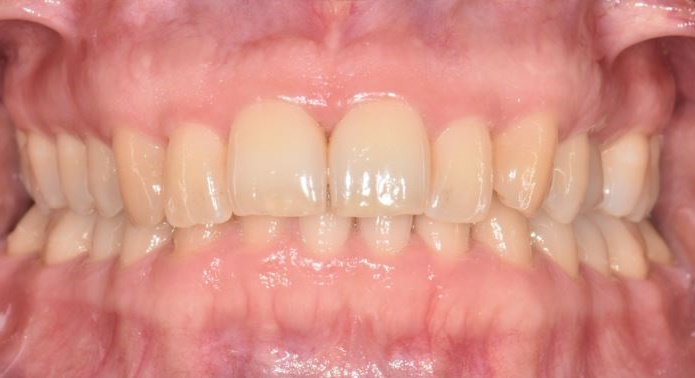
Periodontal health
The clinical presentation and diagnostic criteria that indicate a diagnosis of periodontal health are:
Clinical presentation:
- No visible interdental recession or bone loss.
- Minimal or absent gingival inflammation, gingival tissues stippled.
- No increase in probing pocket depth beyond 3.5 mm.
- Patient may or may not have good plaque biofilm control.
Diagnostic criteria:
- No sites with bone loss >2 mm from cemento-enamel junction due to periodontitis.
- Bleeding on probing at <10% sites.
- Probing pocket depth less than or equal to 3.5 mm
Information on the appropriate data set and clinical management strategy for patients with a diagnosis of periodontal health is presented below.
Required:
- Perform BPE and record results.
- If radiographs have been taken for another purpose (e.g. caries detection), include a radiographic report in the clinical notes confirming absence of bone loss.
- Record presence or absence of risk factors which may influence recall intervals and may indicate the likelihood of periodontal disease developing.
Optional:
- Recording a plaque index is useful to objectively monitor oral hygiene in patients who are at risk of periodontal disease development.
- Recording a bleeding index is useful for monitoring changes in gingival inflammation in patients who are at risk of periodontal disease (gingivitis/periodontitis) development.
- Make a diagnosis, inform the patient and discuss management options.
- Provide Step 1 of treatment (if required):
- Provide advice on risk factor control as indicated (OHI/smoking cessation etc.);
- Provide PMPR as needed to remove calculus that hinders OH and to prevent development of inflammation.
- Arrange recall based on assessment of patient risk of developing gingivitis or periodontitis.
- At the risk-based recall visit:
- Record BPE;
- Check for changes/maintenance of plaque biofilm control and bleeding, using index charts if required;
- Record risk factors;
- Provide advice on risk factor control as indicated (OHI/smoking cessation etc.);
- Provide PMPR as needed to remove calculus hindering OH and prevent inflammation developing.
- Arrange recall based on assessment of patient risk.
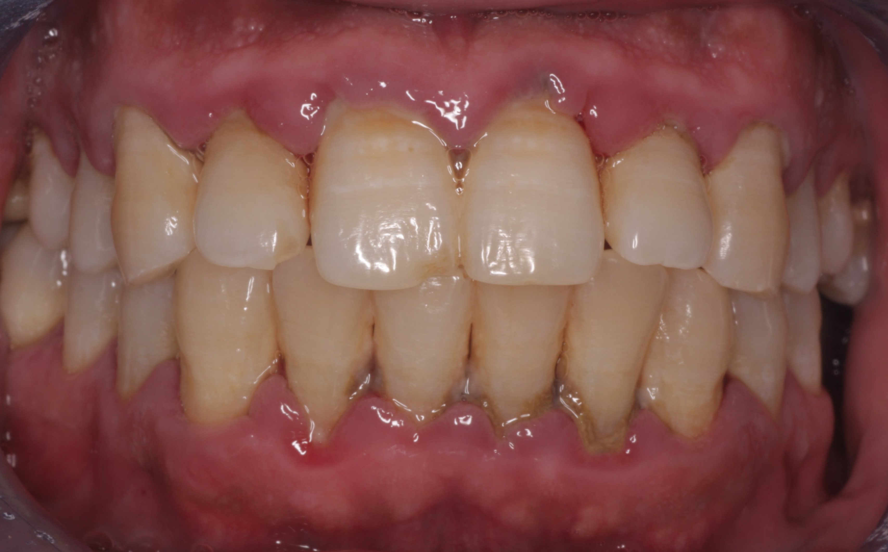
Gingivitis
The clinical presentation and diagnostic criteria that indicate a diagnosis of gingivitis are:
Clinical presentation:
- Red, swollen and inflamed marginal gingival tissue.
- Patient may have halitosis and calculus deposits, both supra- and subgingivally.
- No interdental recession or bone loss.

Diagnostic criteria:
- No sites with bone loss >2 mm from cemento-enamel junction due to periodontitis.
- Bleeding on probing at ≥10% of sites (≥10% of sites for localised gingivitis and >30% sites for generalised gingivitis).
Information on the appropriate data set and clinical management strategy for patients with a diagnosis of gingivitis is presented below.
Required:
- Perform BPE and record results.
- If BPE scores of 3 or 4 are recorded, record a 6-point pocket chart.
- For BPE scores of 3, charting of the affected sextants should be carried out after initial treatment.
- For BPE scores of 4, full mouth charting is required before treatment.
- If radiographs have been taken for another purpose (e.g. caries detection), include a radiographic report in the clinical notes confirming absence of bone loss.
- Record presence or absence of risk factors which may influence recall intervals and may indicate the likelihood of progression to periodontitis.
Optional:
- Recording a plaque index is useful to objectively monitor changes in oral hygiene which influence levels of gingival inflammation.
- Recording a bleeding index is useful for monitoring changes in gingival inflammation over time and also relates to the levels of oral hygiene achieved by the patient.
- Make a diagnosis, inform the patient and discuss treatment options.
- Provide Step 1 of treatment:
- Provide advice on risk factor control as indicated (OHI/smoking cessation etc.);
- Provide PMPR as needed to remove deposits hindering OH, resolve inflammation and reduce probing pocket depth.
- Reassess at subsequent visit:
- Check for resolution of symptoms, bleeding on probing and adequacy of plaque biofilm control;
- Consider objective recording of plaque and bleeding on index teeth.
- If BPE score was 3 before treatment, record a 6-point pocket chart in that/those sextant(s);
- Arrange recall (or further treatment) based on outcome of reassessment and risk of disease progression.
- At risk-based recall visit:
- Record BPE;
- Check for changes/maintenance of plaque biofilm control and bleeding, using index charts if required;
- Monitor gingivitis resolution and absence of progression in PPD and/or recession (or bone loss when radiographs are available);
- Provide advice on risk factor control as indicated (OHI/smoking cessation etc.);
- Provide PMPR as needed to remove calculus hindering OH, resolve inflammation and reduce probing pocket depth;
- Arrange further recall based on a reassessment of patient risk.
Periodontitis
The clinical presentation and diagnostic criteria that indicate a diagnosis of periodontitis are:
Clinical presentation:
- Patient may have red, swollen and inflamed marginal gingival tissue.
- Patient may have halitosis and calculus deposits, both supra- and subgingivally.
- Patient may have drifting or mobile teeth.
- Periodontal pocketing.
- Bleeding on probing.
- Visible interdental recession may be present.
- Bone loss from the CEJ observed on radiographs.
Diagnostic criteria:
- Attachment loss >4 mm at more than 2 non-adjacent sites.
- Bone levels on radiographs >2 mm apically from CEJ.
Information on the appropriate data set and clinical management strategy for patients with a diagnosis of periodontitis is presented below.
- Record risk factors.
- Perform and record BPE.
- Record a 6-point pocket chart (also for all patients with interdental recession).
- Take radiographs of affected teeth and include a radiographic report that describes the amount and extent of bone loss in the clinical notes (see Staging).
- Record a plaque index to objectively monitor changes in oral hygiene which influence levels of gingival inflammation.
- Record a bleeding index to monitor changes in gingival inflammation over time and the level of oral hygiene achieved by the patient.
- Assess current disease status:
- Currently stable: BoP <10%, PPD ≤ 4 mm, no BoP at 4 mm sites;
- Currently in remission: BoP ≥10%, PPD ≤4 mm, no BoP at 4 mm sites;
- Currently unstable: PPD ≥5 mm or BoP at 4 mm sites.
- Make a diagnosis, inform the patient and discuss treatment options.
- Consider prognosis of teeth and the need, or desire, for extractions.
- Provide Steps 1 and 2 of treatment:
- Provide risk factor control advice as indicated (OHI/smoking cessation etc.);
- Provide PMPR as needed to remove deposits on crown and root of the tooth, resolve inflammation and reduce probing pocket depth.
- Reassess at subsequent visit:
- Repeat pocket charting and record levels of oral hygiene and bleeding;
- Consider whether further treatment is required and discuss with patient;
- Consider repeating Steps 1 and 2 of treatment if disease remains active and the patient is engaged with treatment;
- Provide further management if indicated e.g. surgery (Step 3 of treatment);
- Alternatively, refer if case is complex and fits referral criteria;
- If pocket depths and bleeding on probing are sufficiently controlled, move patient to periodontal maintenance, (Step 4 of treatment).
- Provide long term care:
- Arrange risk-based recall visits, likely to be every three months initially and extending to longer recall intervals longer if patient is stable.
- Provide Step 4 of treatment:
- Reassess symptoms, bleeding on probing and plaque biofilm control;
- Provide risk factor control advice as indicated (OHI/smoking cessation etc.);
- Provide PMPR as needed to remove deposits hindering OH, resolve inflammation and reduce probing pocket depth;
- Record a 6-point pocket chart annually for all patients with a diagnosis of periodontitis;
- Consider assessing gingival recession to monitor attachment levels in the long term.